


Sinister Spheres - Extra Footage
Strange Spheres with Unusual Qualities

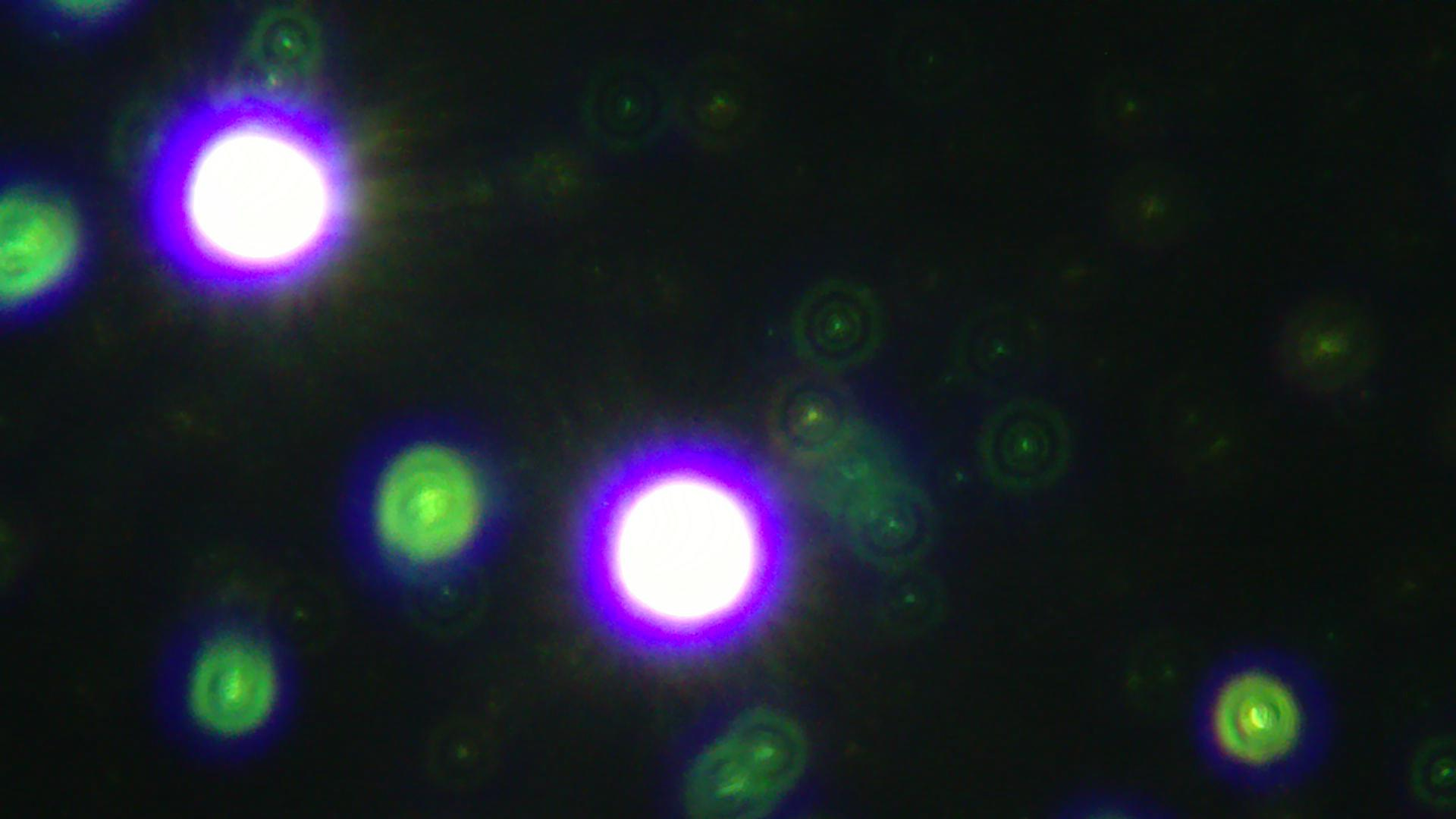
Picture #1 - Papaya (Darkfield) - Glowing Spheres
It looks like this papaya had more than one type of sphere in it. I wonder what each type does.
In 2022, Mat Taylor observed nanostructures in Covid-19 vaccine samples self-assembling in the presence of Wifi, and dissolving in it’s absence. For this reason, I often add Wifi to suspicious structures in samples to see if they change.
While applying EMF to the papaya sample, I was able to catch a sphere as it was dissolving. Enjoy watching it self-destruct in the following video:
Video #1 - Papaya Sample - Sphere Dissolving in Real Time
I looked at some liquid Ivermectin the other day and found some very unusual spheres. Several spheres in the sample seemed to have gel, or another fluid-like substance, around them. See photo below:
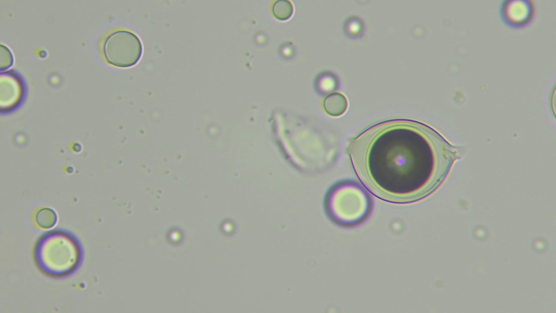
Picture #2 - Liquid Ivermectin Sample - Spherical Nanocarrier With a Fluid-Like Exterior
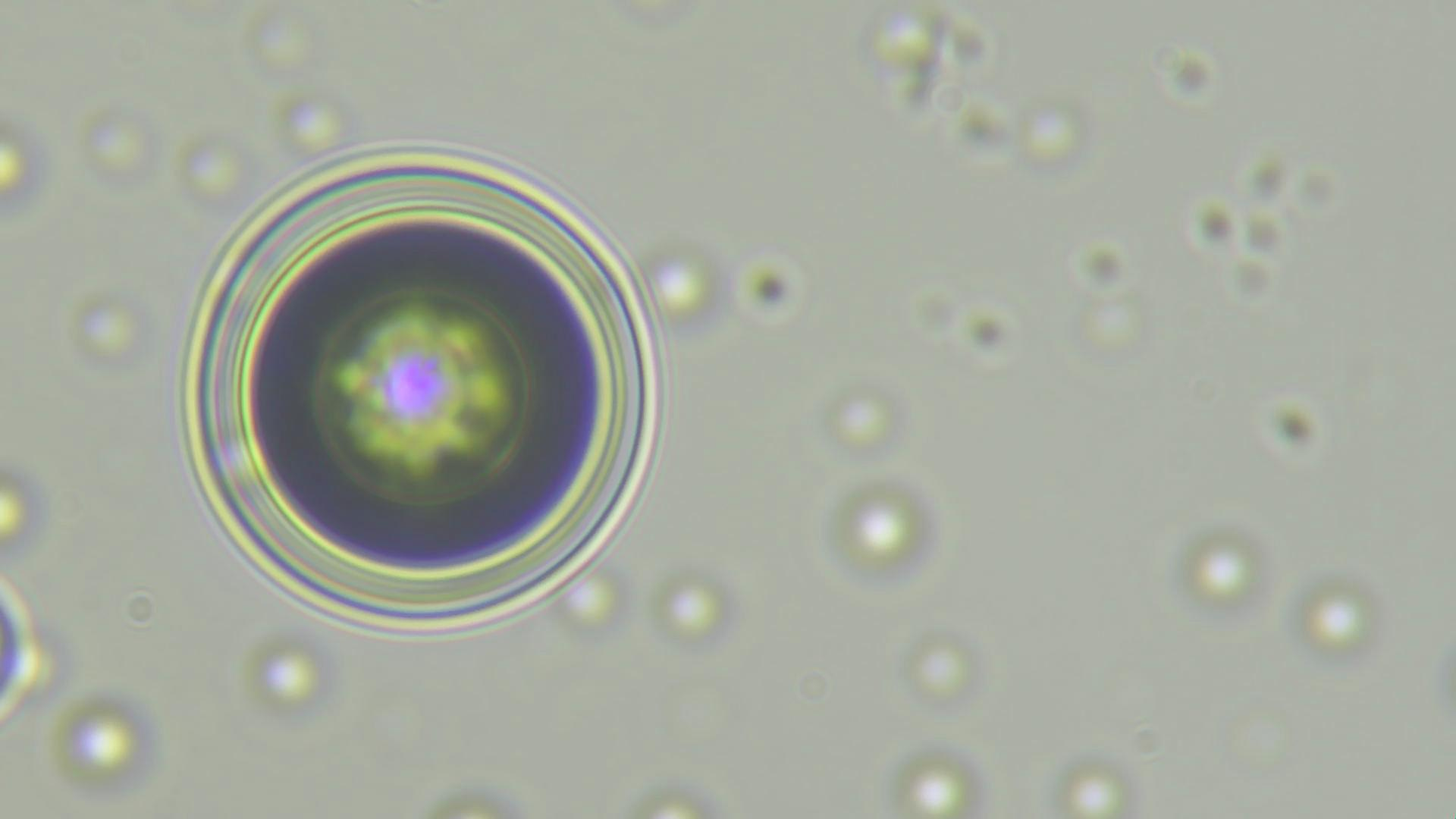
Picture #3 - Liquid Ivermectin Sample - Another Sphere with A Fluid-like Exterior
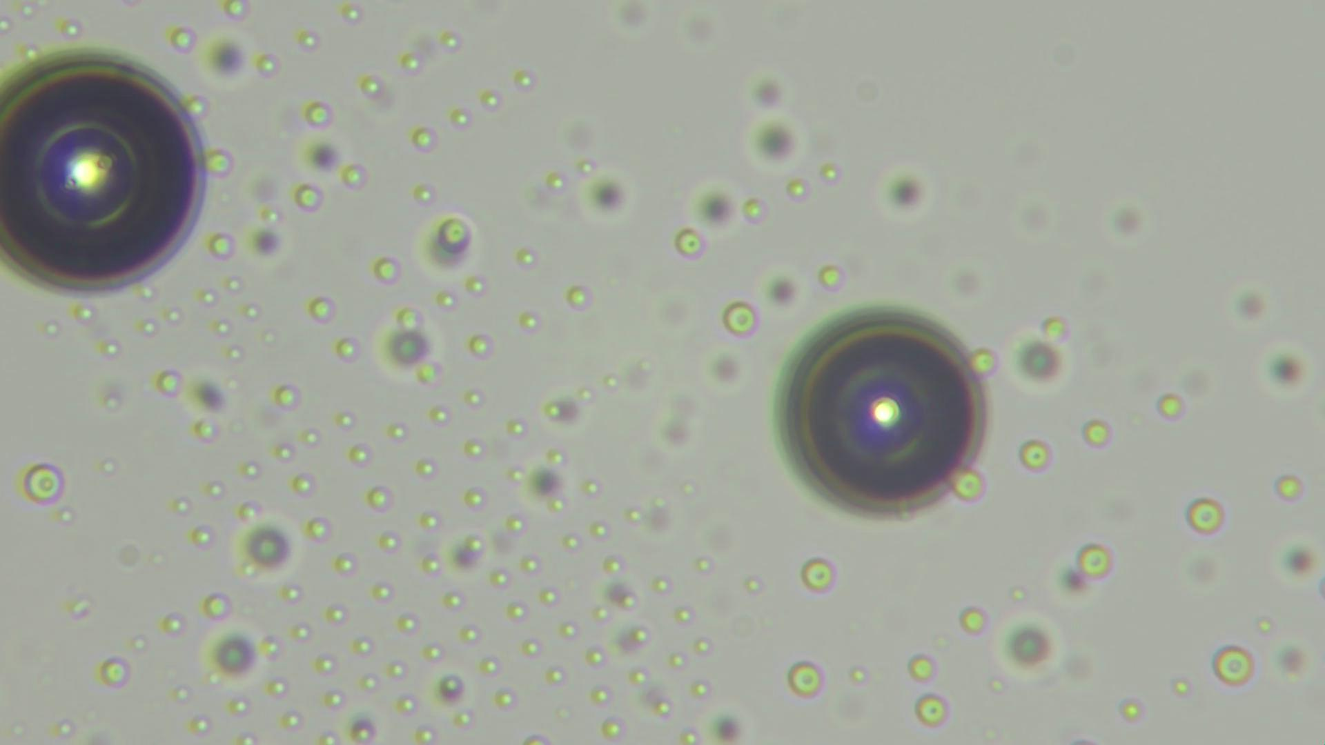
Picture #4 - Same Liquid Ivermectin Sample as Shown Above
Here it looks like there are two large nanocarriers, as well as numerous smaller liquid droplets or spheres arranged in a structured formation.
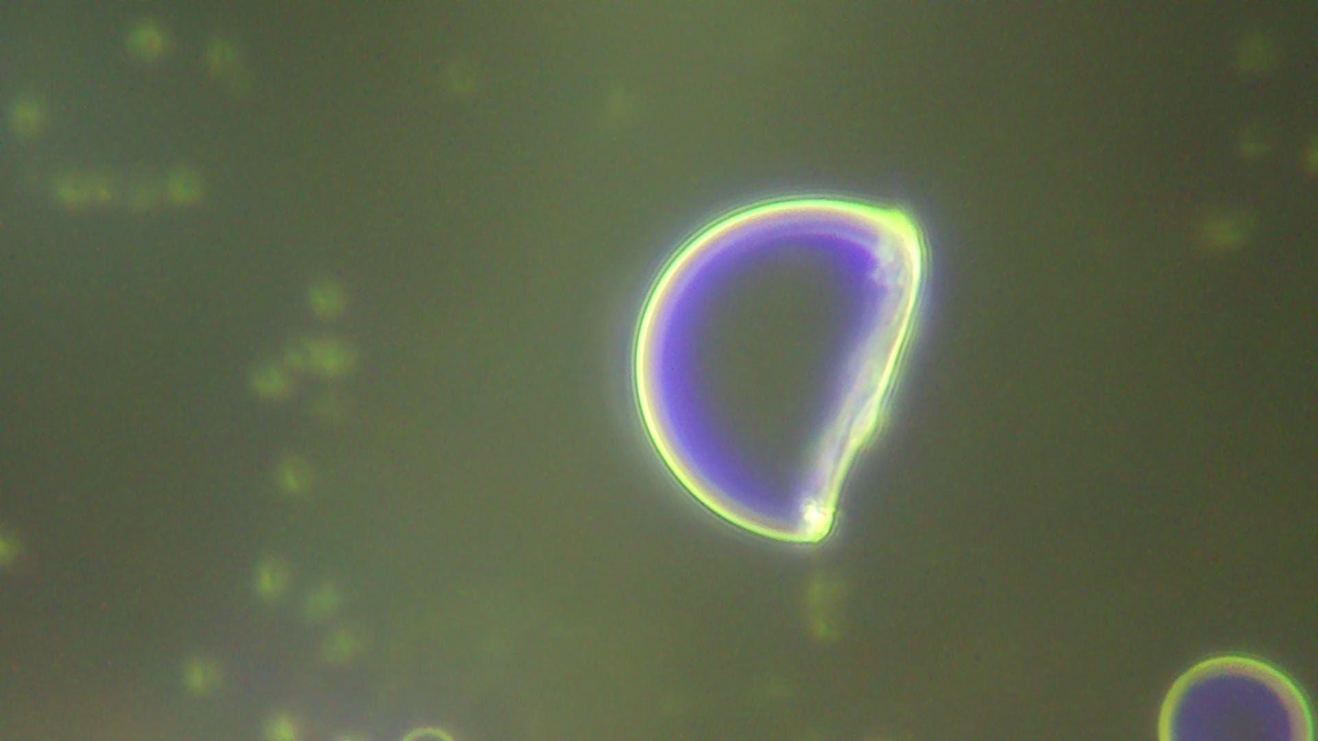
Picture #5 - Liquid Ivermectin Sample - A Sphere with A Bite Taken Out of It?
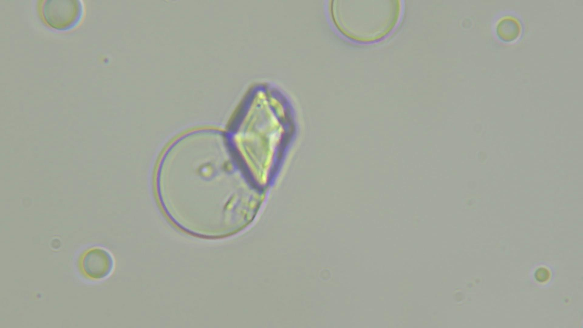
Picture #6 - Liquid Ivermectin Sample - A Sphere with A Pyramid-Shaped Object on Top.
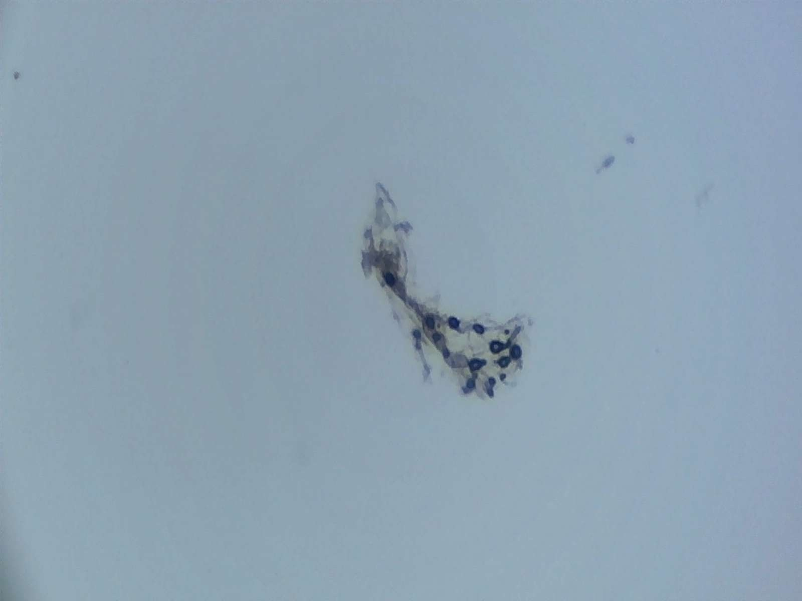
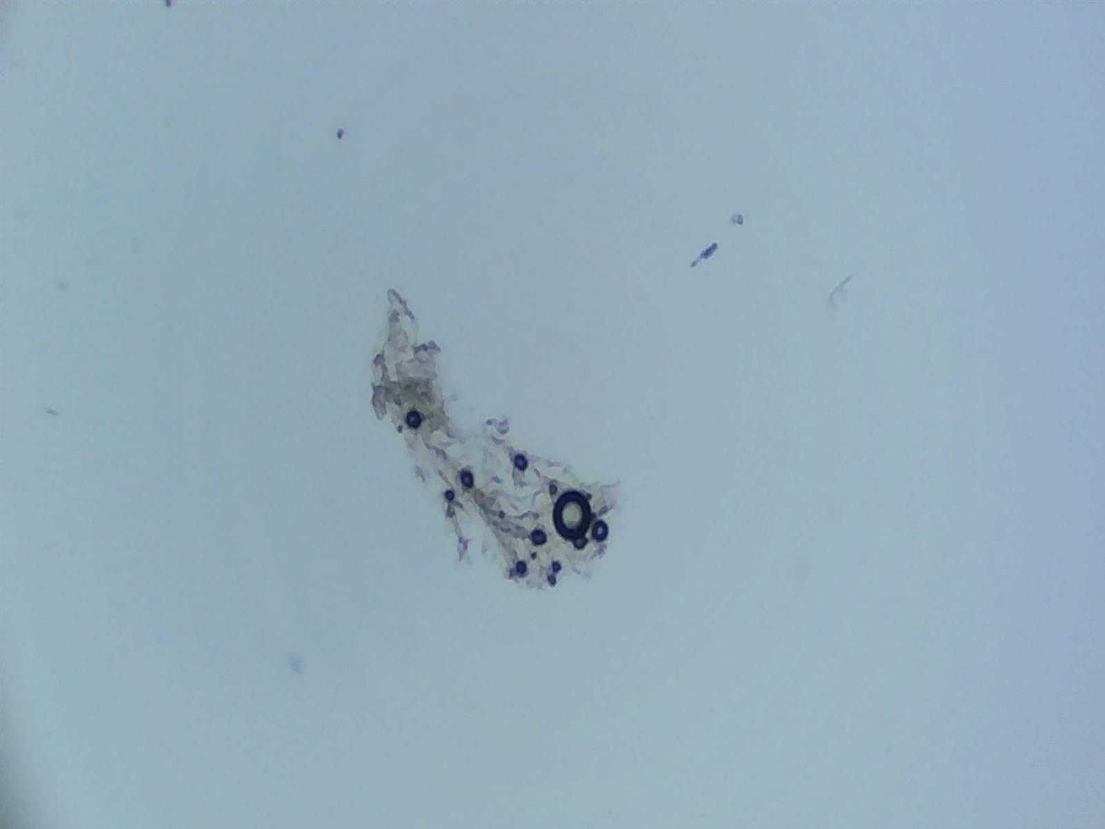
Picture #7 - Liquid EDTA (Brightfield Microscope) - Before adding Wifi.
I added Wifi to the EDTA sample seen above to see what would happen. Pay attention to changes occurring in the sphere with the longest tail on the lower righthand side of the clump.
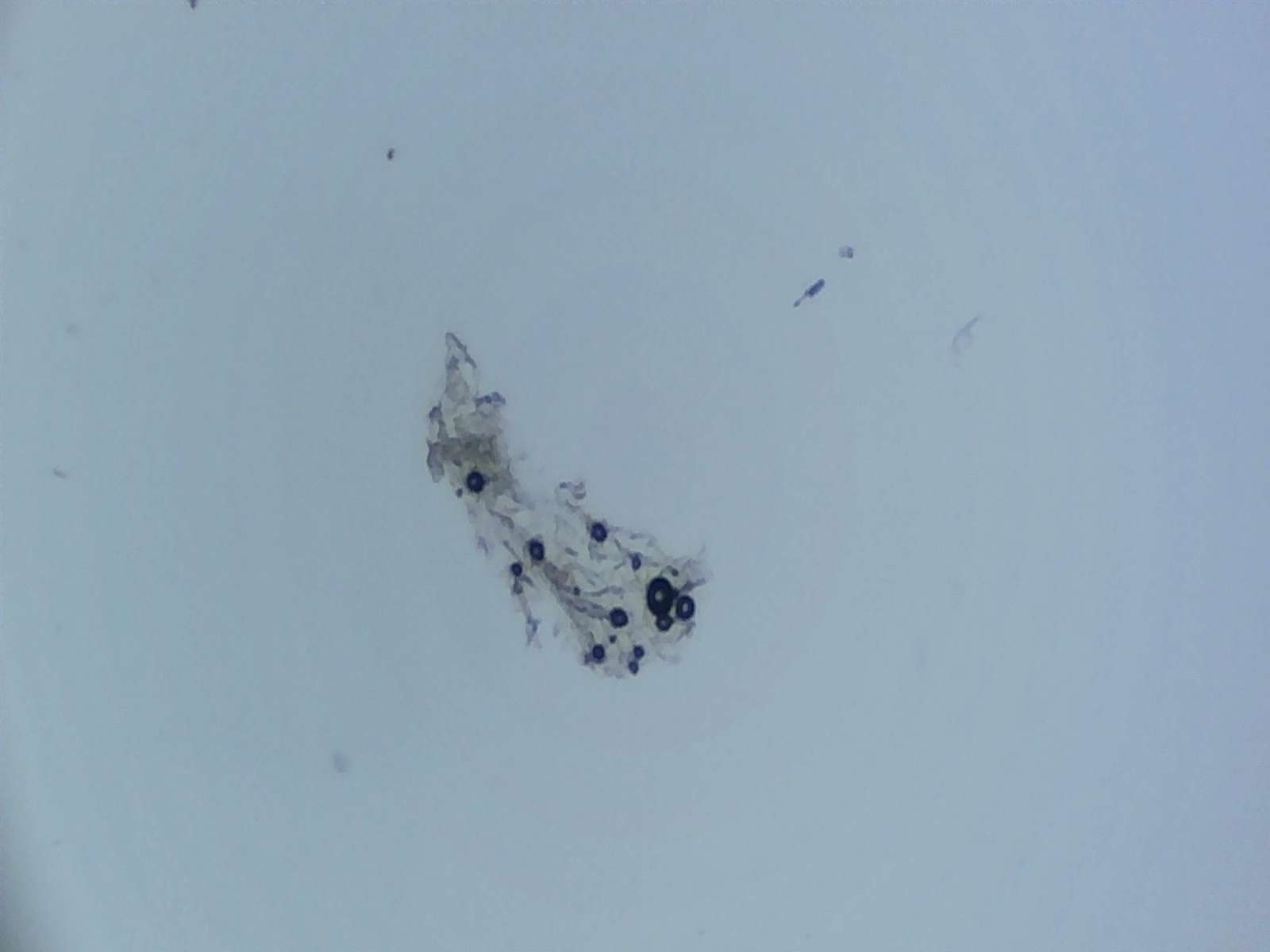
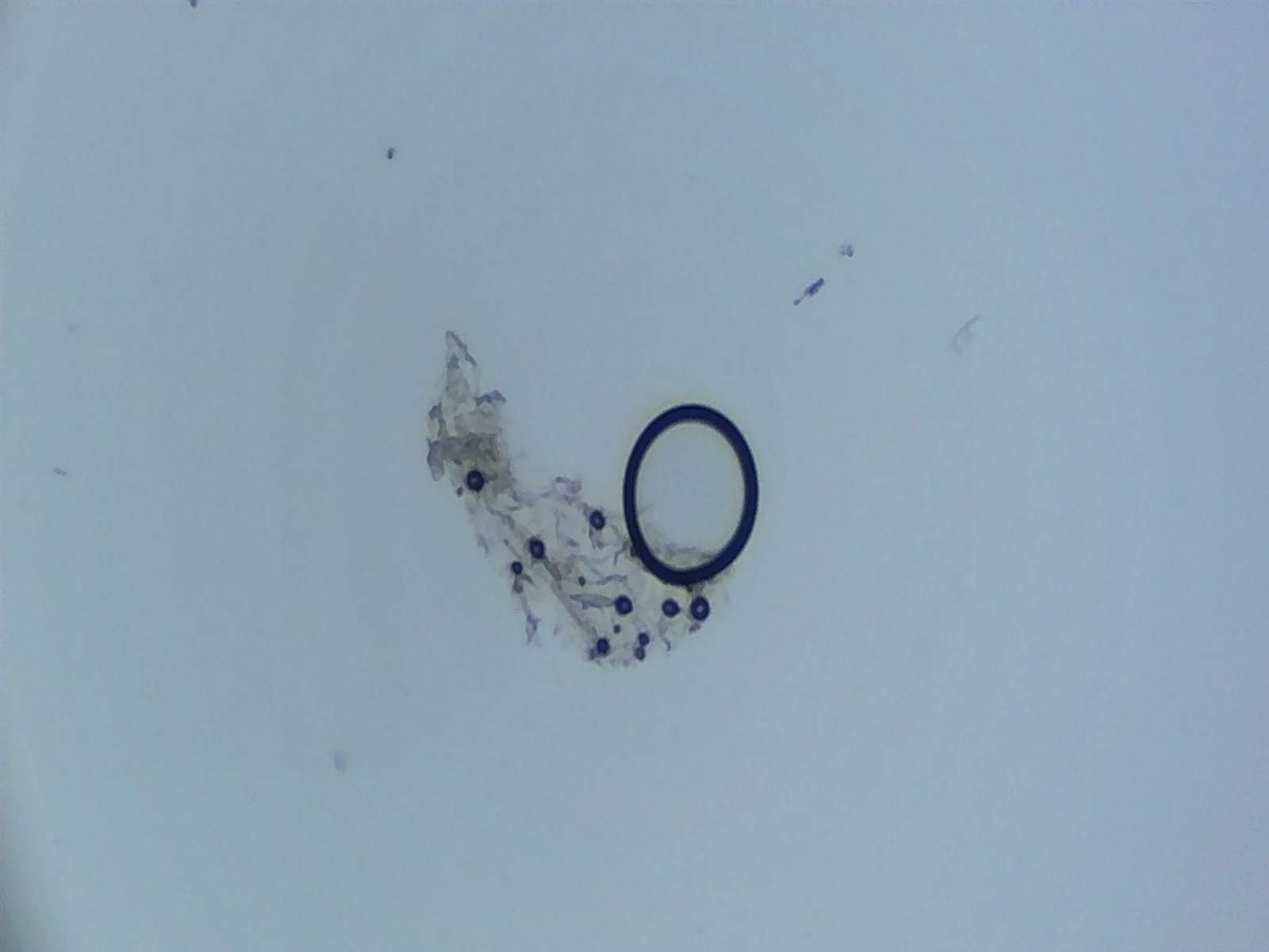
Picture #8 - Liquid EDTA - Same Image After Approximately 90 minutes of Wifi
It looks like some of the spheres seen in Picture #7 have disappeared, moved, or transformed. The one with the tail in Picture #7 looks larger now.
Picture #9 - Liquid EDTA - Same Image After About 2 1/2 Hours of Wifi
Picture #10 - Liquid EDTA After 4 Hours of Wifi - Well… Well… Well… What do we have here?
Do spherical nanocarriers in our bodies respond to Wifi the same way? Could high Wifi exposure cause nanocarriers to expand, pop and release their contents prematurely? Would this lead to higher overall blood toxicity? These are all questions that should be answered.
Picture #11 - Milk Creamer - The Dark Sphere Here Looks Like It’s Dissolving
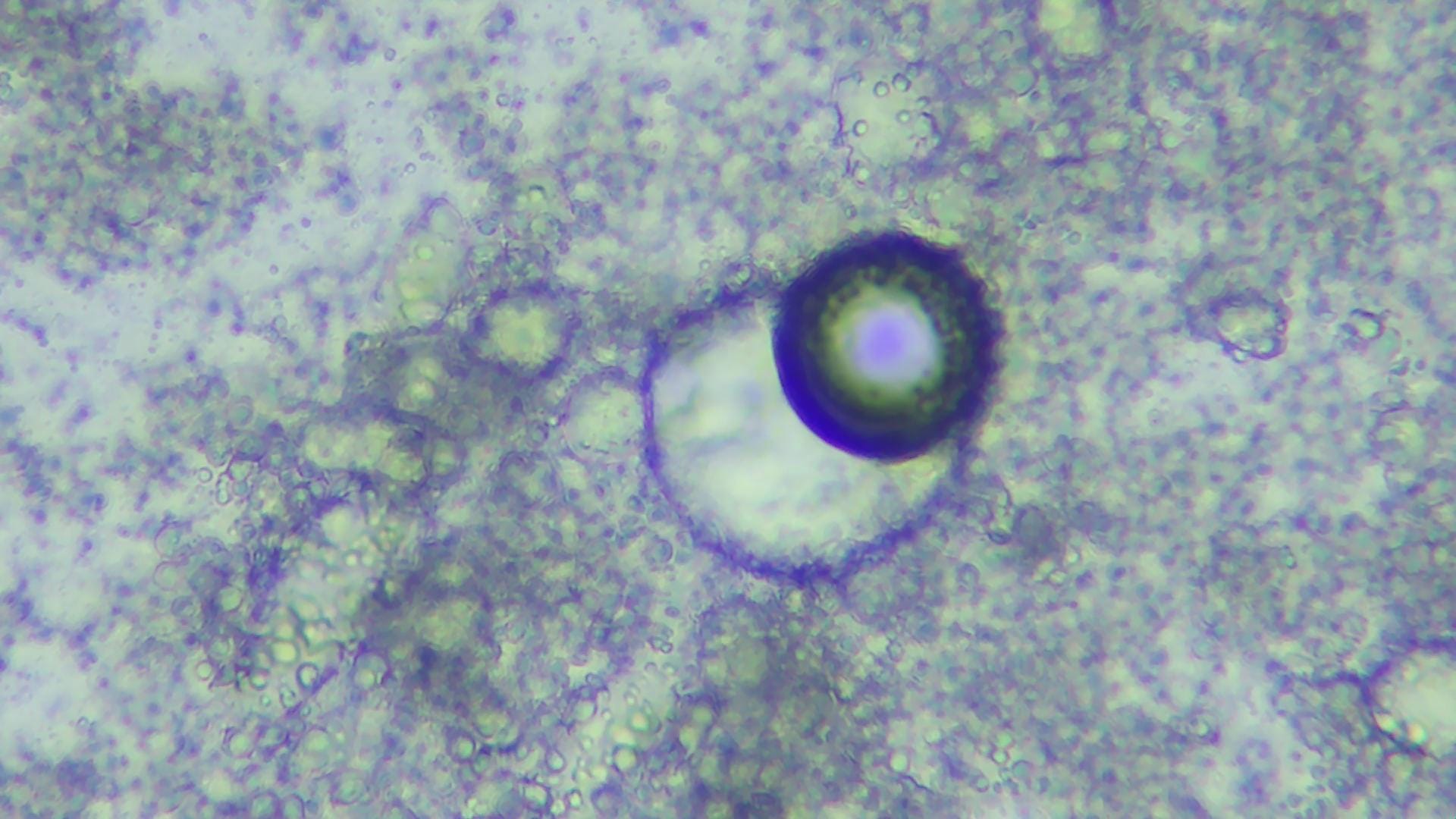
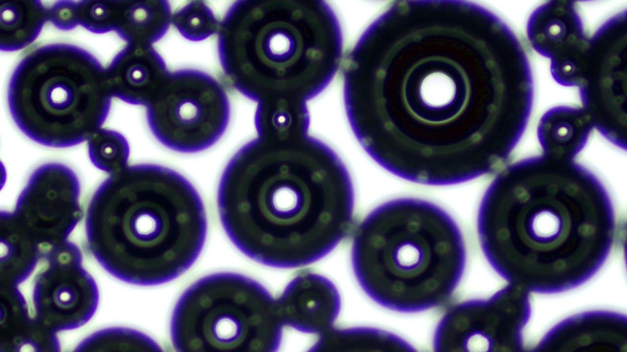
Picture #12 - Papaya Sample
The tiny white dots seen in the second layer of the large sphere above, remind me of a pattern I’ve seen in Ibuprofen gelcaps and beef blood. See below:
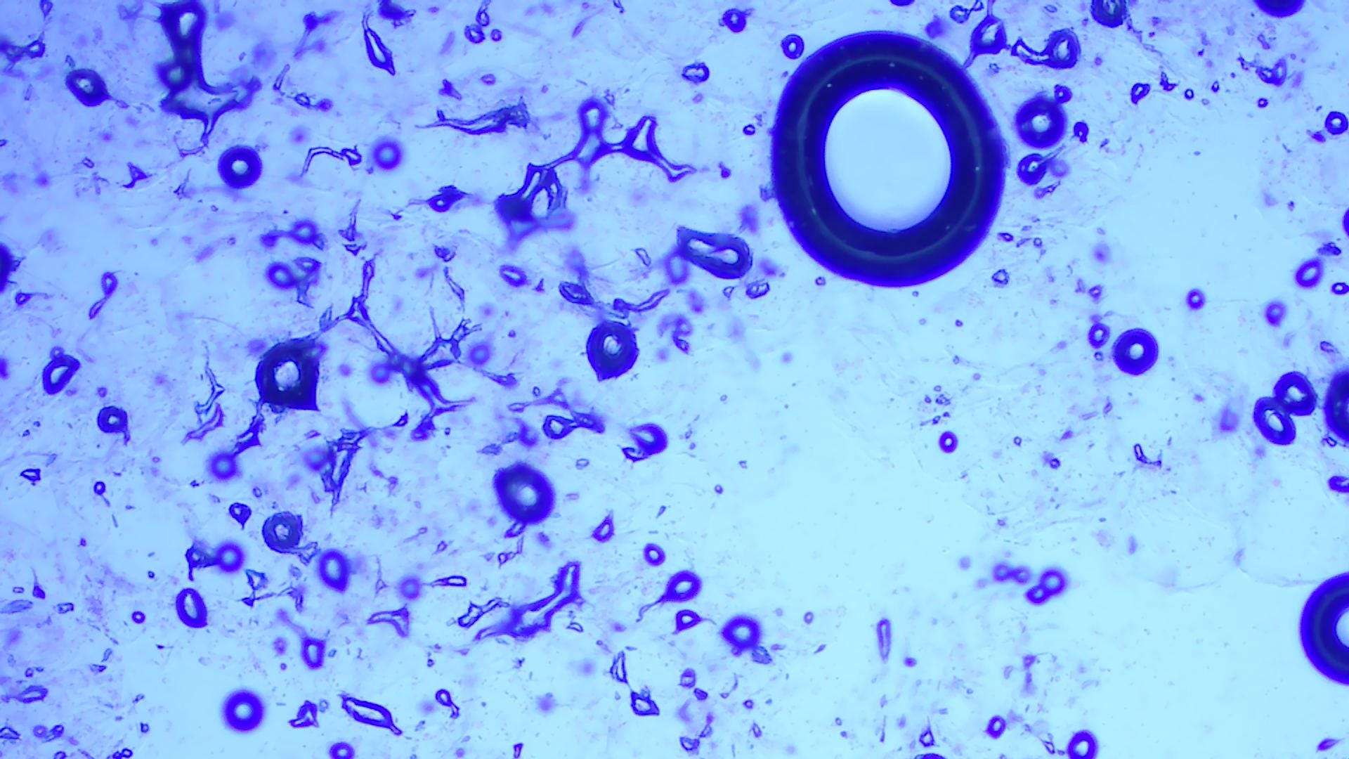
Picture #13 - Ibuprofen Gel cap Sample
This is about scary as it gets. It looks like the lighter-colored spheres inside the black ones facilitate connection. What else do they do? It’s not terribly easy to find the answers to such questions by putting normal search terms into a browser. Fortunately, there’s another way to learn more about nanostructures. Free videos of classes for nanotechnology students posted on YouTube, and other platforms, discuss the purposes of various colors and shapes as part of their coursework.
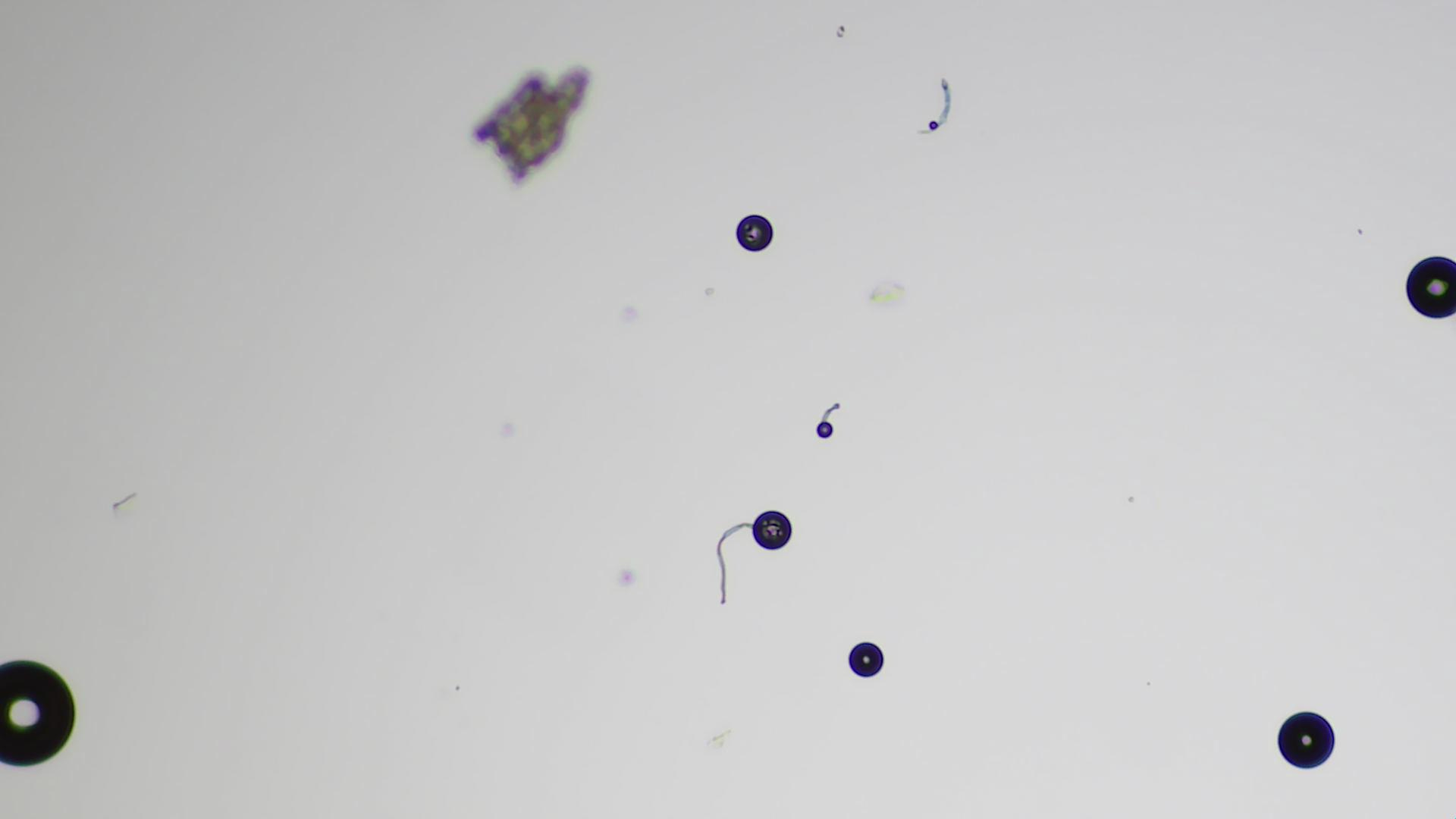
Picture #14 - Spheres with Single Tails(?) in Ear Drops
Drawings of the internal structure of spherical nanocarriers - like liposomes - typically show how phospholipids are used to form bilayers. Phospholipids can have a single tail, but more commonly have two. The trouble is, they are generally too small to be seen with an optical microscope. This leaves the objects with tails above a bit of a mystery.